TL;DR
Skin tags on dogs are small, soft, harmless growths that hang from the skin on a narrow stalk. They are common in older dogs, can appear in any breed, and a dog that gets one often gets more. Most need no treatment. The problem is that several other lumps can look like a skin tag, including ticks, keratin cysts, warts, sebaceous gland growths and mast cell tumors, the most common malignant skin tumor in dogs. The Merck Veterinary Manual recommends a biopsy to confirm a skin tag, and a quick needle sample (fine needle aspirate) agreed with the full lab diagnosis in 90.9% of skin lumps in one study. Our founder Morty has dealt with both ends of this: a pimple-like cyst on one pug, and a 5 cm tumor that had to be surgically removed from another.
What is a skin tag on a dog?
Vets call skin tags acrochordons, soft fibromas or fibrovascular papillomas. According to the Merck Veterinary Manual:
- They are benign and common in older dogs.
- They can develop in any breed, although large breeds may be at increased risk.
- They usually look like a stalk-like growth, often with a wart-like surface.
- They can be single or multiple, and dogs that develop one are prone to develop others over time.
- Removal is optional, but a biopsy is recommended to confirm the diagnosis.
A typical skin tag is skin-colored or slightly darker, soft, painless and moves with the skin. It does not hurt your dog unless it gets caught on a collar or harness, is chewed, or bleeds.
Skin tag or something else?
These are the lumps most often mistaken for a skin tag.
| Lump | What it usually looks like | What to know |
|---|---|---|
| Skin tag | Soft, small, hangs on a stalk, wart-like surface | Benign; often more appear over time |
| Tick | Gray, brown or bluish "bump" stuck to the skin, grows bigger over a few days | Look closely where it meets the skin: a tick has tiny legs. Remove it with fine tweezers |
| Keratin (follicular) cyst | Round bump in the skin, can look like a pimple | Contains a gray, brown or yellowish "cheesy" material. Merck warns not to squeeze it |
| Wart (papilloma) | Rough, cauliflower-like growth | True warts are caused by a virus |
| Sebaceous gland overgrowth or adenoma | Small shiny or crusty bump, often on the head | Common in older dogs; usually benign |
| Lipoma | Soft lump under the skin that moves when touched | A benign fatty tumor, common in older, overweight dogs. See fatty lumps on dogs |
| Mast cell tumor | Raised lump, soft to firm; can look like almost anything | The most common malignant skin tumor in dogs; pugs are predisposed |
| Melanoma | Dark spot, patch or raised lump | Most skin melanomas in dogs are benign, but some are not |
Why you cannot tell by looking
Merck's advice is blunt: skin tumors are so diverse that identifying them "should be left to a veterinarian." Mast cell tumors in particular can mimic harmless lumps such as lipomas, so appearance alone cannot rule them out.
The numbers show why this matters:
- In a German review of 109,616 dog tissue samples, which found 70,966 tumors, the most common after mammary tumors were benign epithelial skin tumors (15.4%) and mast cell tumors (9.7%), followed by histiocytomas (7.0%), soft tissue sarcomas (5.8%) and lipomas (5.8%).
- Among skin tumors sent to pathology labs, 37.1% were malignant in a Portuguese study of 1,185 cases and 56.7% in a Japanese study of 1,435 cases. These are lumps vets were already worried enough to send for testing, so a lump found at home is, on average, less likely to be serious. But they show how often a "lump" turns out to matter.
- The risk of skin tumors rises with age. In the Japanese study it increased significantly from 11 years onward.
The fine needle aspirate: a quick answer
For most lumps, the first test is a fine needle aspirate: the vet pushes a thin needle into the lump, draws out cells and looks at them under a microscope. It is quick and usually needs no sedation. In a study of 243 skin and under-skin lumps from dogs and cats, the needle sample agreed with the full lab diagnosis in 90.9% of cases, and when it said "tumor" it was right 99.4% of the time. If the result is unclear, your vet may suggest a biopsy.
Pugs and mast cell tumors
If you own a pug, take every new lump seriously:
- A US study of veterinary records found pugs had 2.28 times the odds of mast cell tumors compared with other dogs.
- In the same study, 56% of pugs with mast cell tumors had more than one skin tumor, but 94% of the tumors were low to intermediate grade, and the authors concluded mast cell tumors in pugs were relatively benign.
- A study of 168,636 dogs in England also found greater odds of mast cell tumors in pugs, along with Boxers and Staffordshire Bull Terriers.
- Merck notes mast cell tumors occur most often in dogs 8 to 10 years old, most often on the limbs, lower belly and chest.
Mast cell tumors have one more trick: rubbing, scratching or handling one can make it swell, itch and turn red, a reaction vets call Darier's sign. Mast cells release histamine, which causes irritation and itching. That is one more reason not to squeeze a lump you have not had checked.
Two lumps from Morty's pugs
Morty has raised seven pugs, and two of them taught Morty very different lessons about lumps.
The pimple that was a cyst
When one of Morty's pugs was around 5 years old, Morty found a red, raised bump that looked just like a pimple. A vet friend looked at it, shaved the area and said it was fine to simply squeeze it. Morty did it at home: white material came out, just like squeezing a pimple. It healed and never came back.
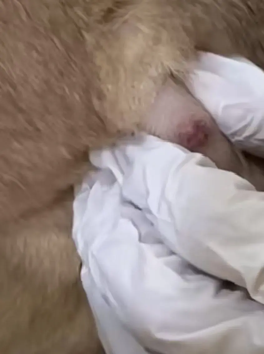
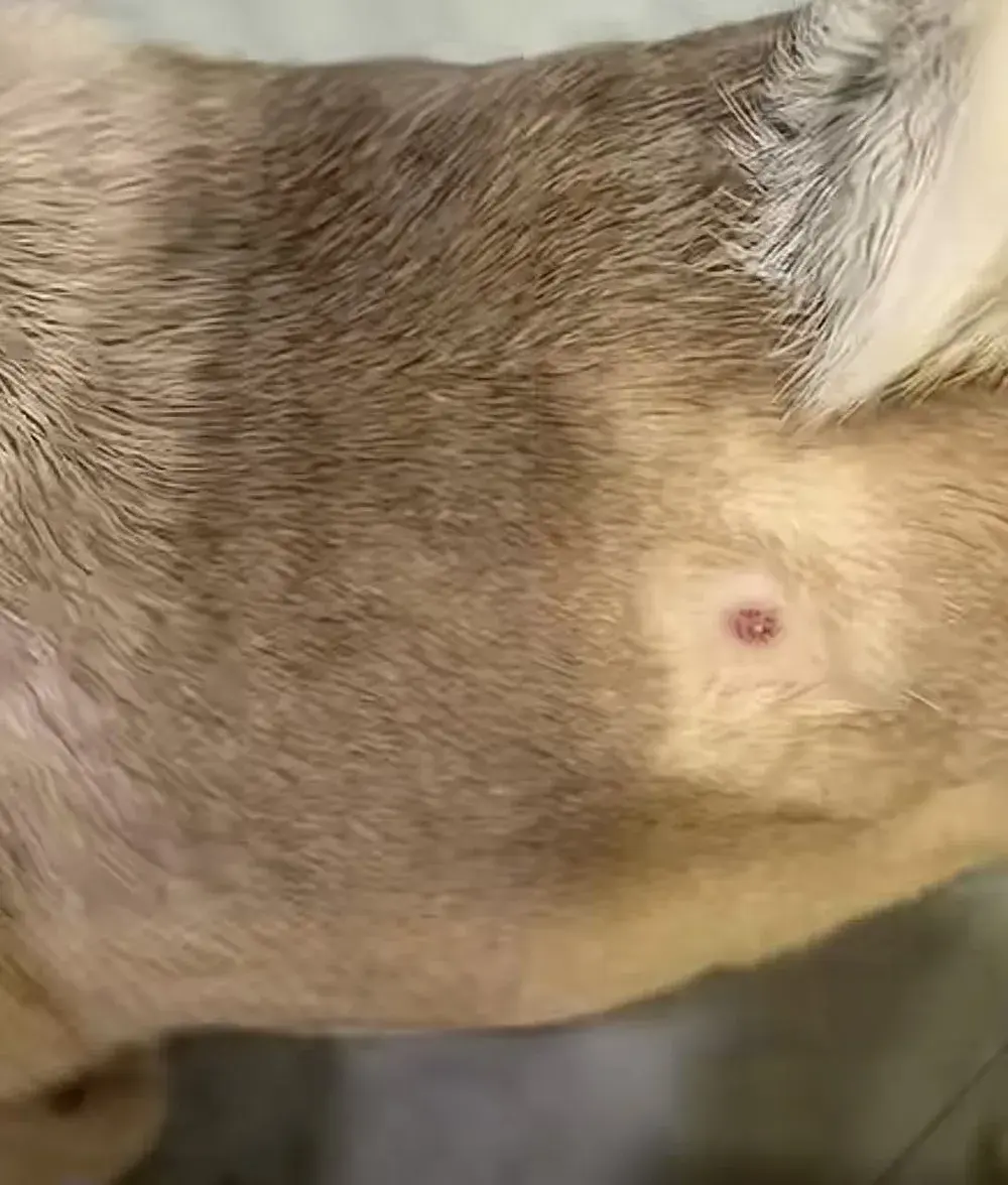
The white, cheesy contents are typical of a keratin cyst, though no test was done. But notice the order of events: a vet looked at the lump first. Without that, Merck advises not squeezing a cyst, because it can push the contents into the surrounding tissue, where the body treats them as a foreign substance and severe inflammation can follow. And if the "pimple" had been a mast cell tumor, squeezing it could have triggered the swelling described above. Have it checked first, every time.
The lump that needed surgery
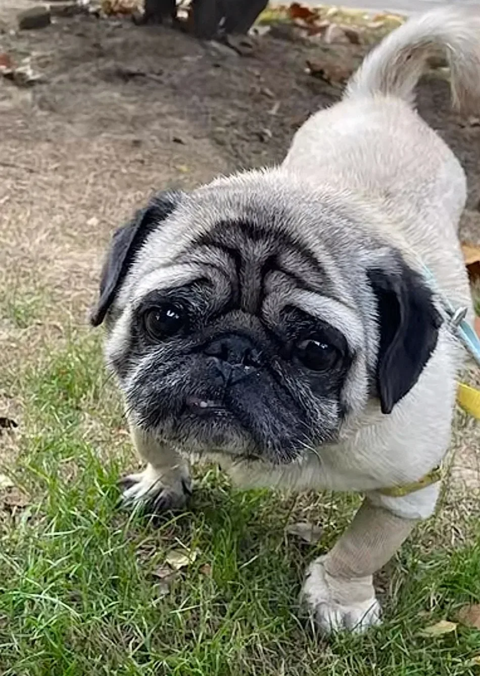
The second story is the one that matters most. When another of Morty's pugs was a little over 9 years old, a tumor grew on its right leg. By the time it was removed it was about 5 cm across. The vet diagnosed a tumor and removed it surgically.
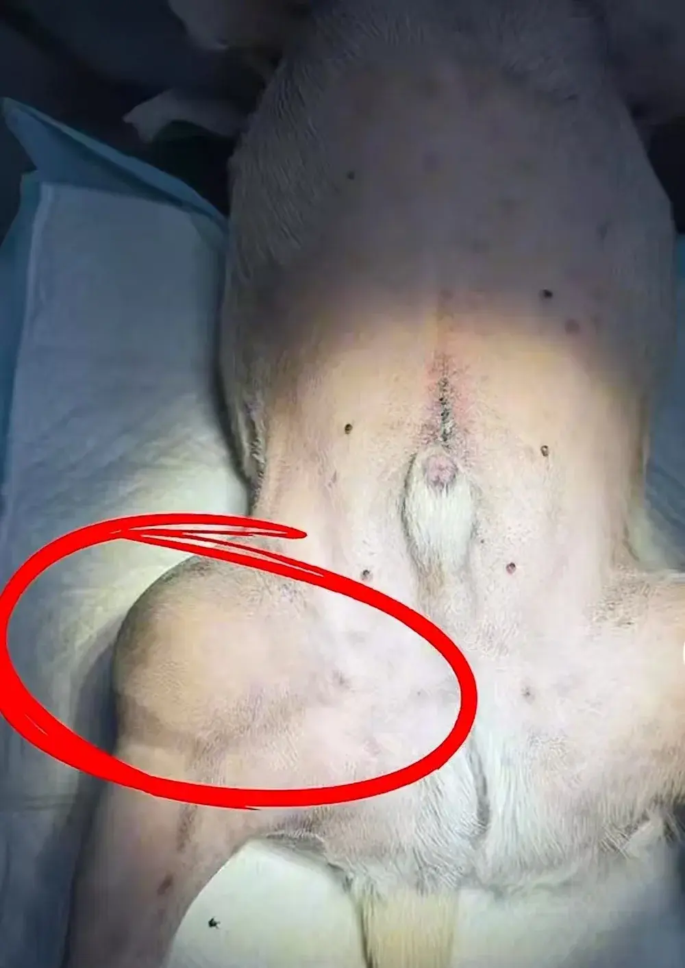
The next photos show the surgery itself.
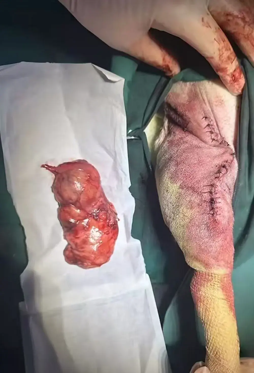
Recovery was faster than Morty expected. After surgery the pug went on an IV drip, and the next day it was already walking normally, just slowly, so Morty took it outside for some fresh air. About three days later, at a routine cleaning and dressing change, the wound was healing well.
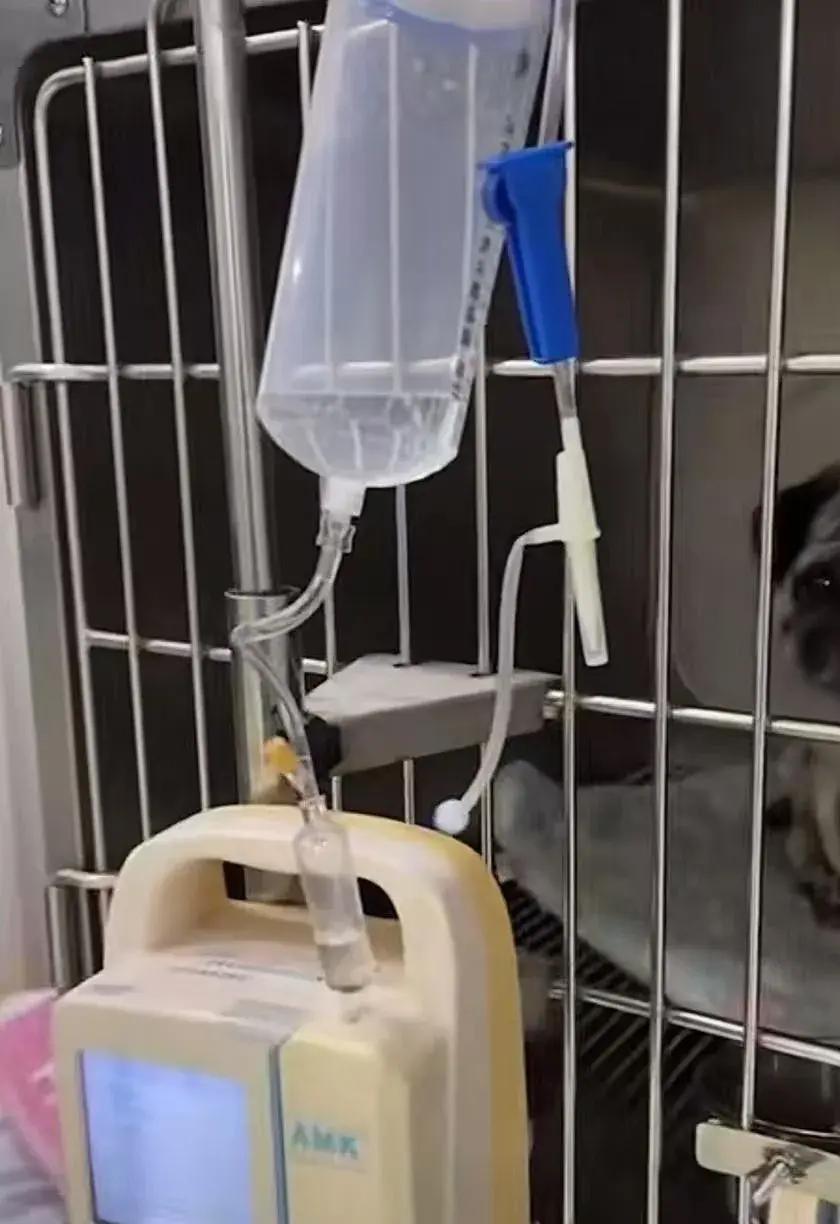
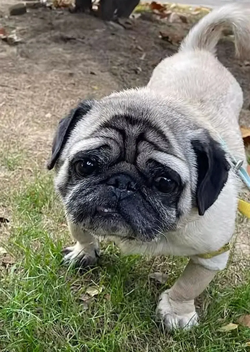
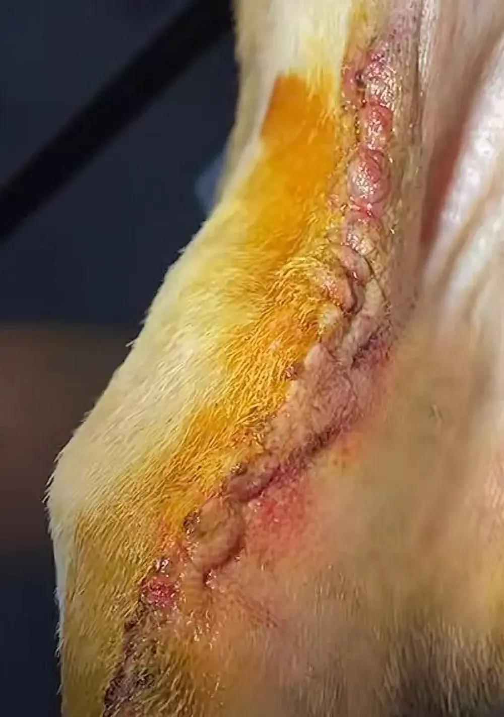
The lesson Morty takes from both: you cannot tell a harmless lump from a dangerous one by looking or feeling, and a lump found early is smaller and easier to remove.
When to see a vet about a lump
Cornell University's Riney Canine Health Center advises a vet visit for any lump that:
- is the size of a pea or larger
- has been there for a month or more
- is growing or changing
- bothers your dog (licking, scratching)
- is red, bleeding, oozing or smelly
Go sooner for any lump on a pug or Boxer, any lump that appears quickly, several new lumps at once, or a lump that swells and shrinks.
Should you remove a skin tag at home?
No. Do not tie off, cut or freeze a skin tag yourself:
- It may not be a skin tag. Removing it at home throws away the chance to test it, and Merck recommends a biopsy to confirm what it is.
- Skin tags have a blood supply. Cutting one can bleed and become infected.
- Squeezing or rubbing can make some lumps worse, as with cysts and mast cell tumors.
If a confirmed skin tag is in the way, catching on a collar or bleeding, your vet can remove it by surgery, electrosurgery or freezing (cryosurgery), sometimes at the same time as another procedure under anesthesia, such as a dental cleaning.
Keep a lump map
Many lumps are found during grooming. Once a month, run your hands slowly over your dog's whole body: head, neck, chest, belly, legs, tail and between the toes. For each lump, write down:
- Where it is (for example "left chest, two fingers behind the elbow")
- How big it is, measured with a ruler or compared with a pea, grape or walnut
- What it feels like: soft or firm, moves or fixed, on the skin or under it
- The date
Take a photo with a coin next to it. If a lump grows or changes between checks, book a vet visit. See senior dog grooming for a full at-home check, and pug lifespan for the other health problems to watch in pugs.
FAQ
When should you worry about a skin tag on a dog?
When it is growing, changing color, bleeding, red or oozing, bothering your dog, or larger than a pea, or when you are not sure it is a skin tag at all. Several lumps, including mast cell tumors, can look like skin tags, so a vet check with a needle sample is the only way to be sure.
How do I get rid of my dog's skin tag?
Usually you do not need to. If it bothers your dog, catches on a collar or bleeds, your vet can remove it by surgery, electrosurgery or freezing. Do not cut or tie it off at home.
Why did my dog suddenly get skin tags?
Skin tags are common in older dogs, and a dog that gets one is prone to develop more over time. Several new lumps appearing at once should still be checked by a vet, especially in pugs, which often develop multiple mast cell tumors.
Can a skin tag look like a tick?
Yes. An attached tick can look like a gray or brown skin tag. Look closely where it meets the skin: a tick has tiny legs there, and it gets bigger over a few days as it feeds. Remove ticks with fine tweezers, gripping close to the skin and pulling steadily.
How do I know if my dog's skin tag is cancerous?
You cannot tell by looking. A vet takes a fine needle aspirate, which in one study matched the full lab diagnosis in 90.9% of skin lumps, or a biopsy.
Can I squeeze a lump on my dog that looks like a pimple?
Not before a vet has seen it. Squeezing a keratin cyst can push its contents into the surrounding tissue and cause severe inflammation, and squeezing a mast cell tumor can make it swell and redden.
References
- Merck Veterinary Manual, Pet Owner Version. Villalobos AE. Tumors of the Skin in Dogs. Last updated 2026.
- Merck Veterinary Manual. Connective Tissue Tumors in Animals. 2026.
- Aupperle-Lellbach H, et al. Tumour incidence in dogs in Germany: a retrospective analysis of 109,616 histopathological diagnoses (2014-2019). Journal of Comparative Pathology. 2022. PMID: 36116890
- Martins AL, et al. Retrospective study of canine cutaneous tumors submitted to a diagnostic pathology laboratory in Northern Portugal (2014-2020). Canine Medicine and Genetics. 2022. PMID: 35216632
- Kok MK, et al. Retrospective study of canine cutaneous tumors in Japan, 2008-2017. Journal of Veterinary Medical Science. 2019. PMID: 31257236
- Ghisleni G, et al. Correlation between fine-needle aspiration cytology and histopathology in the evaluation of cutaneous and subcutaneous masses from dogs and cats. Veterinary Clinical Pathology. 2006. PMID: 16511787
- McNiel EA, Prink AL, O'Brien TD. Evaluation of risk and clinical outcome of mast cell tumours in pug dogs. Veterinary and Comparative Oncology. 2006. PMID: 19754824
- Shoop SJ, et al. Prevalence and risk factors for mast cell tumours in dogs in England. Canine Genetics and Epidemiology. 2015. PMID: 26401329
- Willmann M, et al. Proposed diagnostic criteria and classification of canine mast cell neoplasms: a consensus proposal. Frontiers in Veterinary Science. 2021. PMID: 34957277
- Cornell University College of Veterinary Medicine, Riney Canine Health Center. Skin Lumps and Bumps. 2023.
For individual health questions, your veterinarian remains the best partner.